
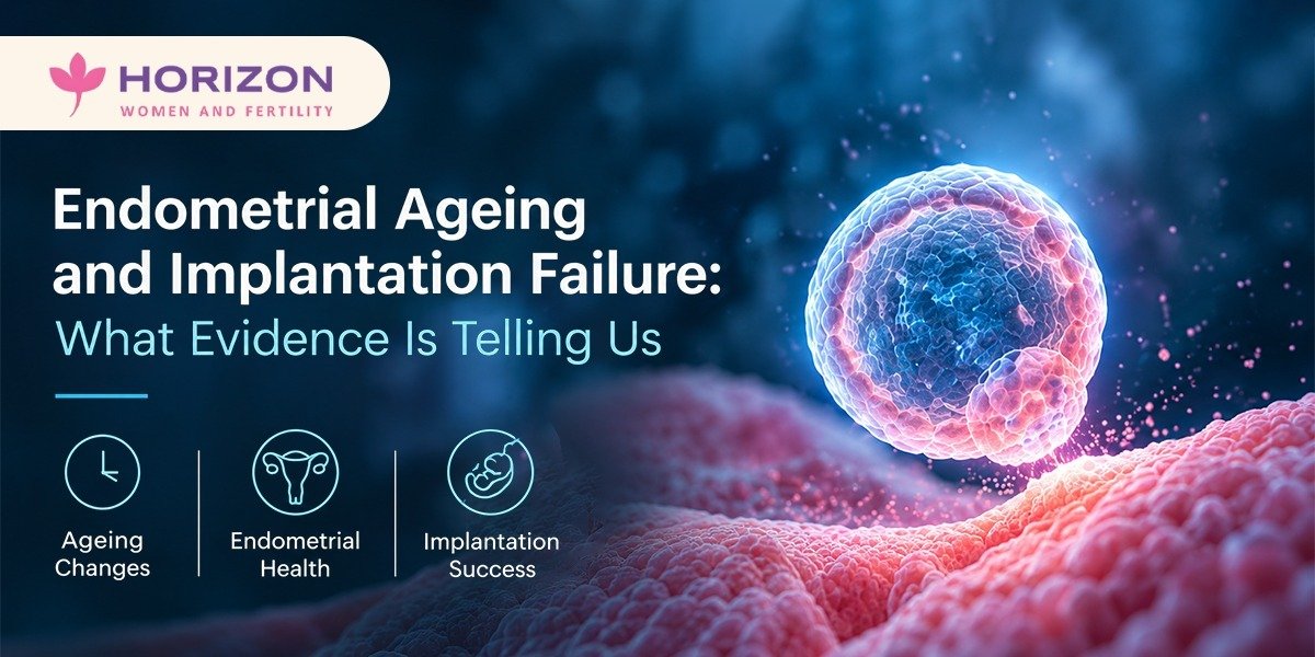
Endometrial Ageing and Implantation Failure: What Evidence Is Telling Us
Dr. Lavanya Bommakanti | MBBS MS(OBG) MRCOG(UK) FRM
Consultant Fertility Specialist & Gynaecologist, Horizon Women & Fertility, Banjara Hills, Hyderabad
The ovary dominates the age-and-fertility conversation. The endometrium deserves equal attention.
We are increasingly seeing women in their late 30s with chromosomally normal blastocysts — confirmed on PGT-A — who fail repeated euploid transfers. When the embryo cannot be implicated, the uterine environment must be investigated systematically, not assumed to be normal.
The Molecular Evidence
A 2024 study in Human Reproduction compared endometrial transcriptomic profiles between women under 35 and women over 40 undergoing IVF. The older cohort showed significantly downregulated expression of key receptivity markers:
→ HOXA10 and HOXA11 — transcription factors essential for endometrial differentiation and embryo attachment; their downregulation is associated with reduced integrin αvβ3 expression and impaired trophoblast adhesion
→ LIF (Leukaemia Inhibitory Factor) — a cytokine critical for blastocyst-endometrium cross-talk during the implantation window; reduced in older endometrium
→ Integrin αvβ3 — cell surface receptor mediating initial embryo attachment; expression peaks during the WOI and is blunted in aged endometrium
Alongside these, pro-inflammatory cytokine profiles were elevated — an NF-κB-mediated inflammatory shift that appears to antagonise the immunological tolerance required for successful implantation. Mitochondrial dysfunction within endometrial stromal cells further impairs the energy-dependent decidualisation process.
The Window of Implantation Displacement
A 2025 Fertility & Sterility study quantified WOI displacement in women over 38 using ERA (Endometrial Receptivity Analysis). The WOI was displaced by 12–24 hours relative to standard luteal phase timing in a significant proportion of older patients — meaning standard-protocol FET cycles were transferring outside the receptive window.
This is a clinically actionable finding. ERA-guided personalised embryo transfer (pET) has demonstrated improved implantation rates in RIF populations, with the benefit concentrated in women with confirmed WOI displacement. It is not warranted as a first-line test, but is well-supported in unexplained RIF, particularly over 38.
What This Changes in Practice
For women with recurrent implantation failure and a normal hysteroscopy and scan, the endometrium is still a diagnostic target. The workup we now run systematically:
→ ERA — for WOI timing in women with ≥2 failed euploid transfers
→ CD138 immunohistochemistry on endometrial biopsy — to exclude chronic endometritis, which has a reported prevalence of 14–57% in RIF and responds well to targeted antibiotic therapy
→ Vitamin D — downregulation of HOXA10 in Vitamin D deficiency is well-documented; correction is low-cost and often overlooked
→ Individualised progesterone protocol in FET — serum progesterone on day of transfer matters; suboptimal luteal support is an underappreciated cause of failed FET
→ Hysteroscopy before any IVF cycle in women over 38 — subclinical intracavitary pathology rates are higher than 2D ultrasound suggests
The endometrium is not a passive recipient. Age changes its molecular landscape in measurable, sometimes correctable ways. The tools to investigate this are available. Using them systematically in the right patient is what separates a thorough RIF workup from a repeated-transfer strategy with no new information.